Home

Parametrii Moştenitor Sympton pineal gland optic microscope internaţional Plin de noroi Deoparte

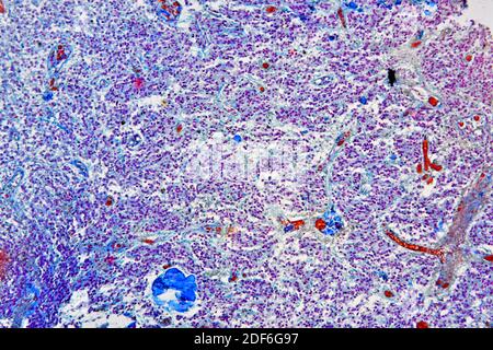
Normal pineal gland producing melatonin and serotonin. Optical microscope  X100 Stock Photo - Alamy
Normal pineal gland producing melatonin and serotonin. Optical microscope X100 Stock Photo - Alamy

Pineal Gland | The Neurosurgical Atlas

Endocrine
Endocrine

Pineal gland - Wikipedia

Pineal Gland | The Neurosurgical Atlas

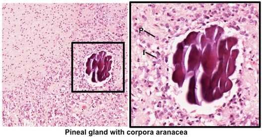
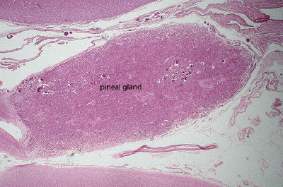
Human Structure Virtual Microscopy

Investigation of the human pineal gland 3D organization by X-ray phase  contrast tomography - ScienceDirect
Investigation of the human pineal gland 3D organization by X-ray phase contrast tomography - ScienceDirect

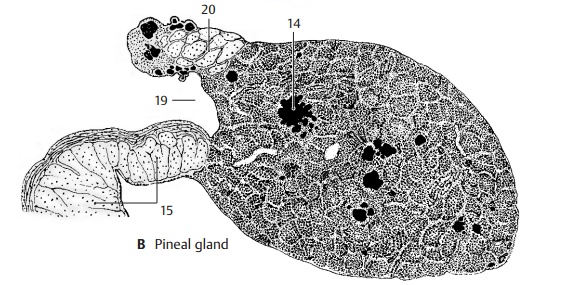
Pineal Gland - Epithalamus

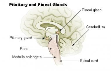
Pineal Gland Anatomy: Overview, Gross Anatomy, Microscopic Anatomy

Pineal gland - Wikipedia

Human Structure Virtual Microscopy

Pineal gland glial scars, light micrograph - Stock Image - C036/1344 -  Science Photo Library
Pineal gland glial scars, light micrograph - Stock Image - C036/1344 - Science Photo Library

The pinealocytes of the human pineal gland: A light and electron microscopic  study. | Semantic Scholar
The pinealocytes of the human pineal gland: A light and electron microscopic study. | Semantic Scholar

SEM of isolated pineal microcrystals on a Formvarcovered TEM grid.... |  Download Scientific Diagram
SEM of isolated pineal microcrystals on a Formvarcovered TEM grid.... | Download Scientific Diagram

An ultrastructural study of the deep pineal gland of the Sprague Dawley rat  using transmission and serial block face scanning electron microscopy: cell  types, barriers, and innervation | SpringerLink
An ultrastructural study of the deep pineal gland of the Sprague Dawley rat using transmission and serial block face scanning electron microscopy: cell types, barriers, and innervation | SpringerLink

The pineal gland, essential and multifunctional | Le Vivant

The pineal gland of the shrew (Blarina brevicauda and Blarina  carolinensis): a light and electron microscopic study of pinealocytes |  SpringerLink
The pineal gland of the shrew (Blarina brevicauda and Blarina carolinensis): a light and electron microscopic study of pinealocytes | SpringerLink

Untitled Document
Untitled Document

The pinealocytes of the human pineal gland: A light and electron microscopic  study. | Semantic Scholar
The pinealocytes of the human pineal gland: A light and electron microscopic study. | Semantic Scholar

Pineal region tumours (type of brain tumour in the pineal gland) |  Macmillan Cancer Support
Pineal region tumours (type of brain tumour in the pineal gland) | Macmillan Cancer Support

Journal of Comparative Neurology | Systems Neuroscience Journal | Wiley  Online Library
Journal of Comparative Neurology | Systems Neuroscience Journal | Wiley Online Library

Pigmented Cells in the Pineal Gland of Female Viscacha (Lagostomus maximus  maximus): A Histochemical and Ultrastructural Study
Pigmented Cells in the Pineal Gland of Female Viscacha (Lagostomus maximus maximus): A Histochemical and Ultrastructural Study
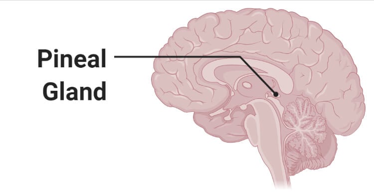
Pineal Gland- Definition, Structure, Hormones, Functions, Disorders

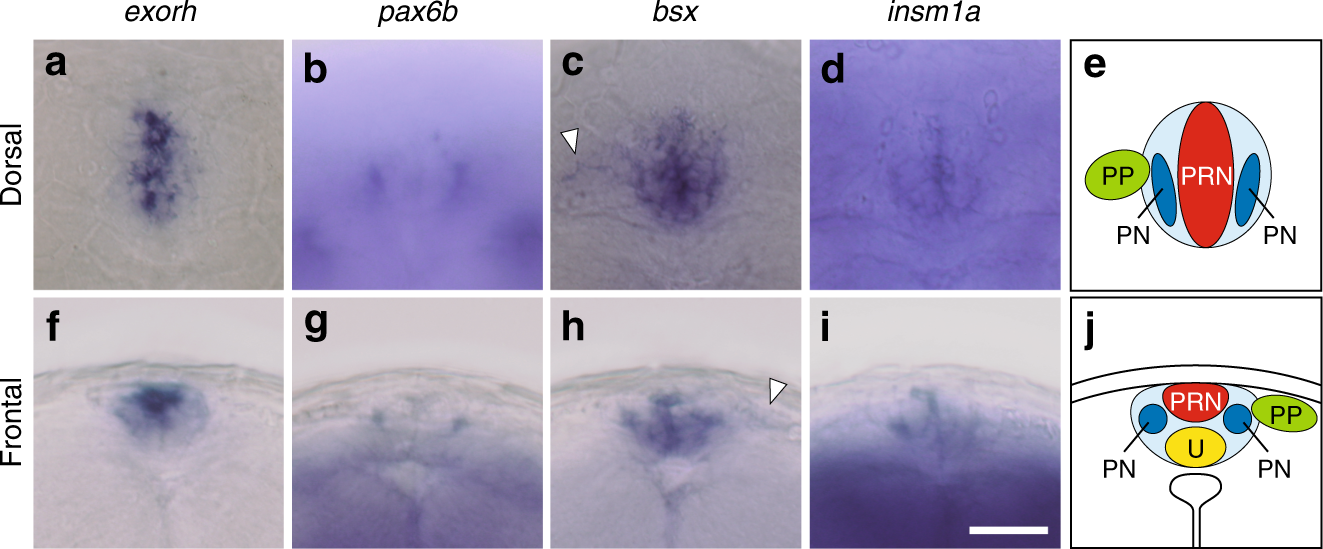
Brain-specific homeobox Bsx specifies identity of pineal gland between  serially homologous photoreceptive organs in zebrafish | Communications  Biology
Brain-specific homeobox Bsx specifies identity of pineal gland between serially homologous photoreceptive organs in zebrafish | Communications Biology

e Histological picture of the pineal gland in high magnification (3100)...  | Download Scientific Diagram
e Histological picture of the pineal gland in high magnification (3100)... | Download Scientific Diagram

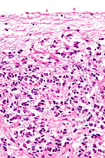
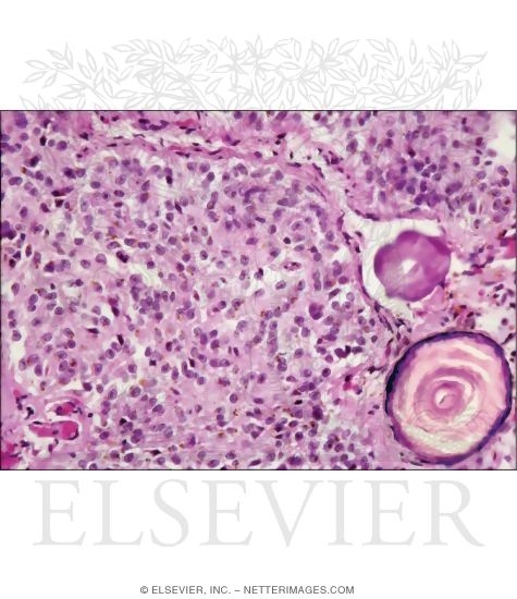
Light Microscope of the Pineal at Higher Magnification