Home

Miere Concediere de lemn electron microscope synapse Jucăuş Loc de noapte dupa aceea

Synapse Dr.Jastrow's electron microscopic atlas

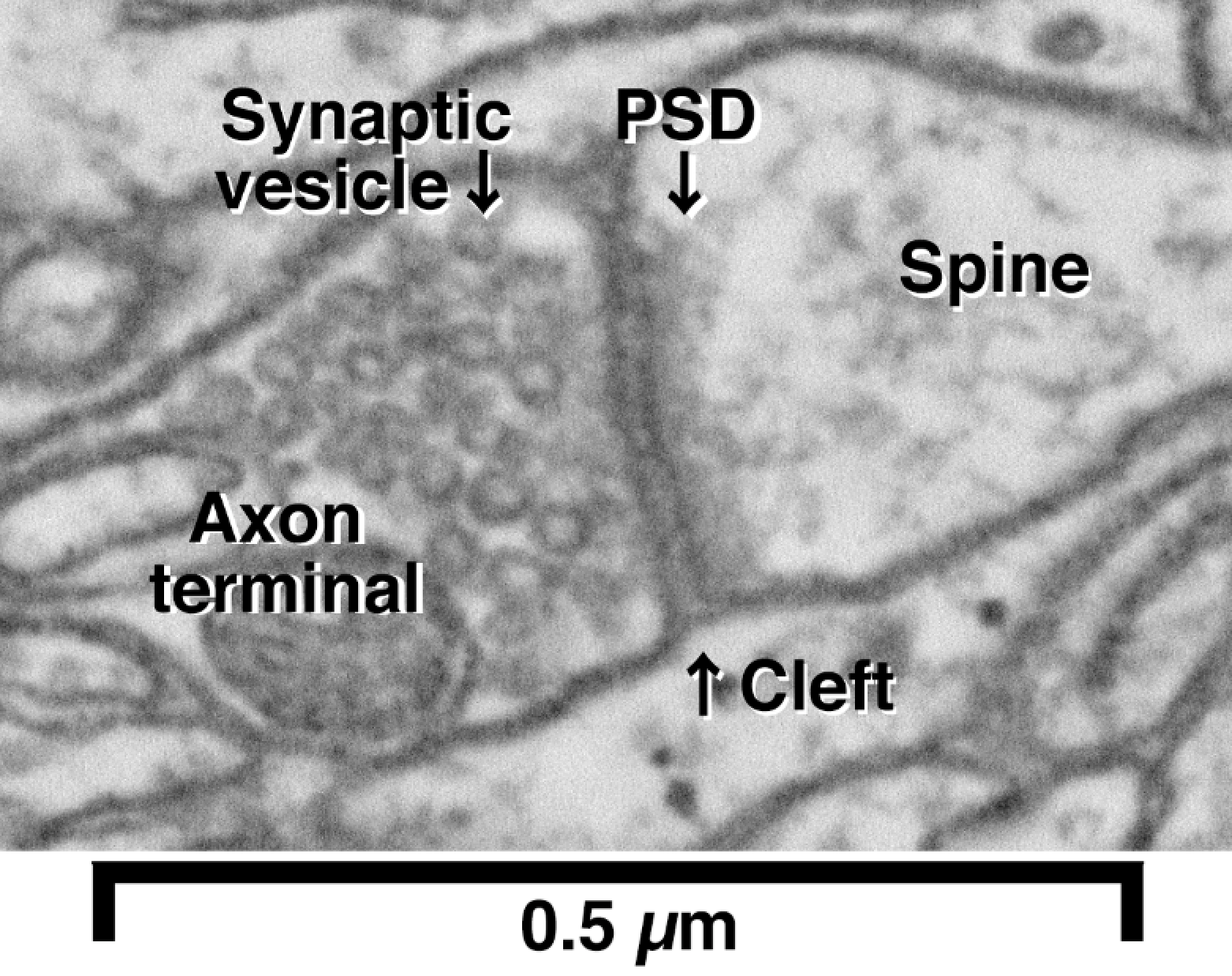
High Magnification Electron Micrograph of a Typical Synapse In the Brain

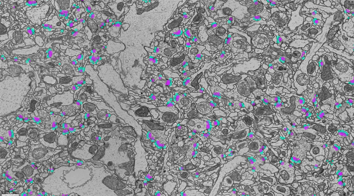
Automatic Detection and Neurotransmitter Prediction of Synapses in Electron  Microscopy | bioRxiv
Automatic Detection and Neurotransmitter Prediction of Synapses in Electron Microscopy | bioRxiv

Probabilistic fluorescence-based synapse detection | PLOS Computational  Biology
Probabilistic fluorescence-based synapse detection | PLOS Computational Biology

Online competition sparks synapse-detection tool | Janelia Research Campus

synapse
synapse

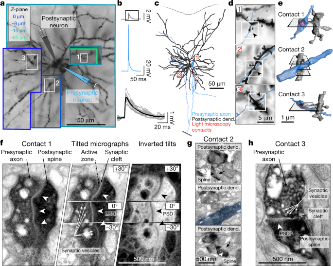
Structure and function of a neocortical synapse | Nature

Synapse Dr.Jastrow's electron microscopic atlas

Early electron microscopic observations of synaptic structures in the  cerebral cortex: a view of the contributions made by Georg
Early electron microscopic observations of synaptic structures in the cerebral cortex: a view of the contributions made by Georg

Reconstructing brain circuits - ZEISS Microscopy Developer Community

Early electron microscopic observations of synaptic structures in the  cerebral cortex: a view of the contributions made by Georg
Early electron microscopic observations of synaptic structures in the cerebral cortex: a view of the contributions made by Georg

Electron micrographs of synapses in layers 1a and 1b in mouse piriform... |  Download Scientific Diagram
Electron micrographs of synapses in layers 1a and 1b in mouse piriform... | Download Scientific Diagram
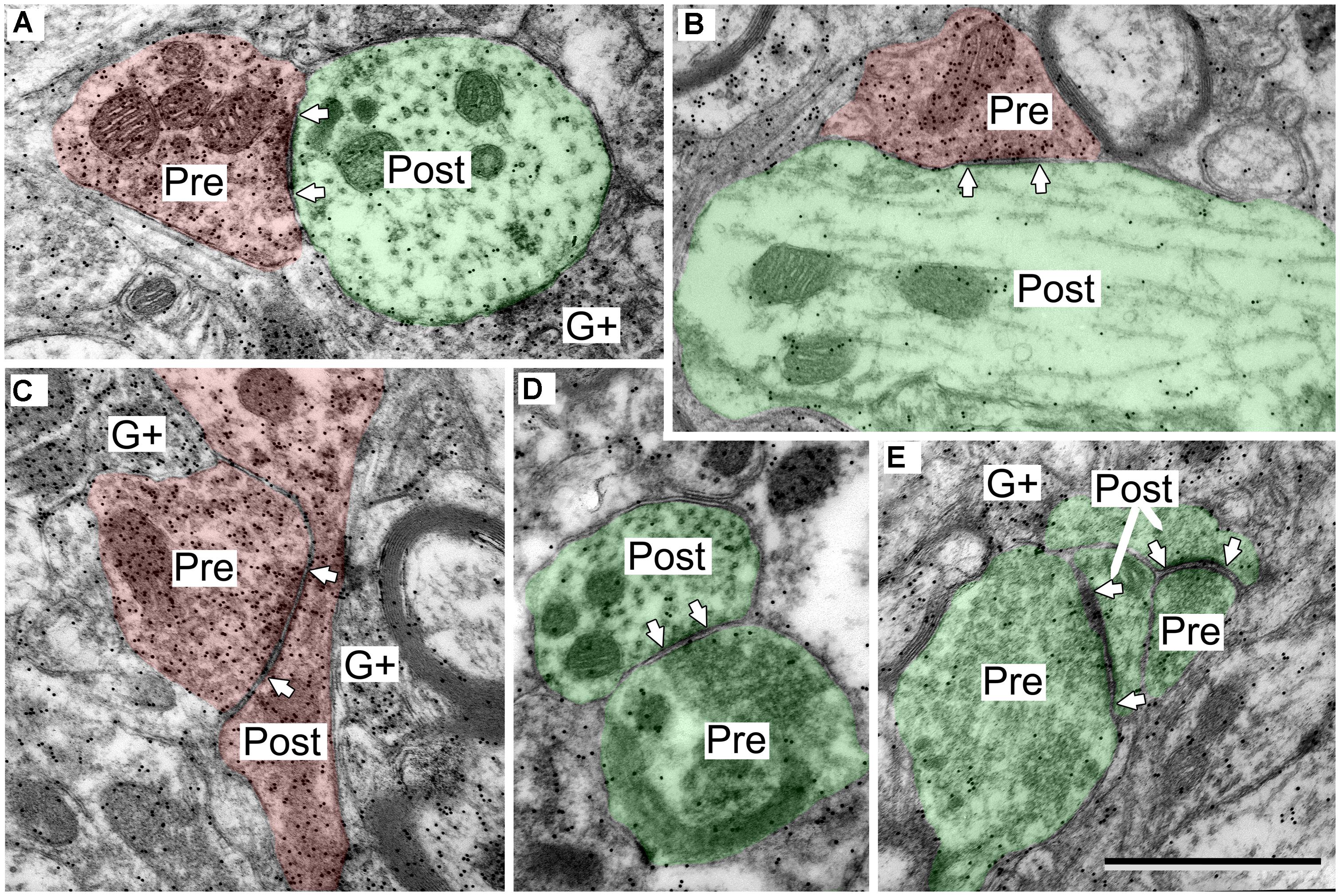
Frontiers | Ultrastructural characterization of GABAergic and excitatory  synapses in the inferior colliculus
Frontiers | Ultrastructural characterization of GABAergic and excitatory synapses in the inferior colliculus

3D Electron Microscopy Study of Synaptic Organization of the Normal Human  Transentorhinal Cortex and Its Possible Alterations in Alzheimer's Disease  | eNeuro
3D Electron Microscopy Study of Synaptic Organization of the Normal Human Transentorhinal Cortex and Its Possible Alterations in Alzheimer's Disease | eNeuro

Synapse EM This EM image reveals a synapse between an axon and dendrite. |  Macro and micro, Medical illustration, Plasma membrane
Synapse EM This EM image reveals a synapse between an axon and dendrite. | Macro and micro, Medical illustration, Plasma membrane
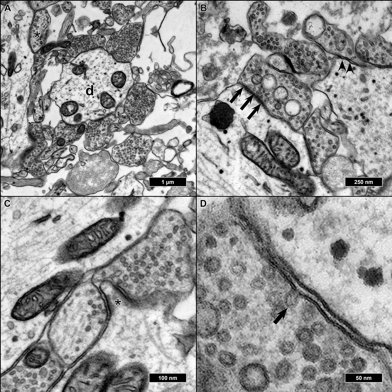
IJMS | Free Full-Text | Visualizing the Synaptic and Cellular  Ultrastructure in Neurons Differentiated from Human Induced Neural Stem  Cells—An Optimized Protocol | HTML
IJMS | Free Full-Text | Visualizing the Synaptic and Cellular Ultrastructure in Neurons Differentiated from Human Induced Neural Stem Cells—An Optimized Protocol | HTML

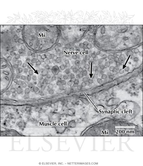
Electron Micrograph of Synaptic Vesicles at a Neuromuscular Junction

Functional Electron Microscopy, “Flash and Freeze,” of  Identified Cortical Synapses in Acute Brain Slices
Functional Electron Microscopy, “Flash and Freeze,” of Identified Cortical Synapses in Acute Brain Slices
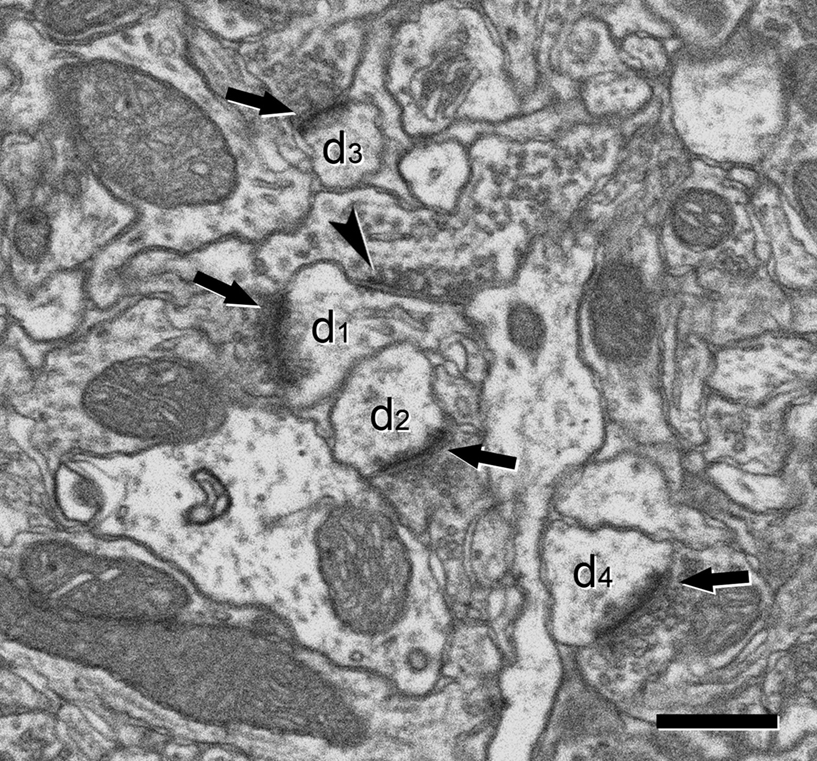
Synapses of Stratum Radiatum | SynapseWeb

Frontiers | Counting synapses using FIB/SEM microscopy: a true revolution  for ultrastructural volume reconstruction
Frontiers | Counting synapses using FIB/SEM microscopy: a true revolution for ultrastructural volume reconstruction

1. A neuronal synapse (upper) Electron microscopy of a synapse: The... |  Download Scientific Diagram
1. A neuronal synapse (upper) Electron microscopy of a synapse: The... | Download Scientific Diagram

A high magnification image of synapse obtained by electron microscopy |  Okinawa Institute of Science and Technology Graduate University OIST
A high magnification image of synapse obtained by electron microscopy | Okinawa Institute of Science and Technology Graduate University OIST

Neuroscience for Kids - Electron Microscopy

Investigating Synapses in Brain Slices with Enhanced Functional Electron  Microscopy | Science Lab | Leica Microsystems
Investigating Synapses in Brain Slices with Enhanced Functional Electron Microscopy | Science Lab | Leica Microsystems

Automatic Detection and Neurotransmitter Prediction of Synapses in Electron  Microscopy | bioRxiv
Automatic Detection and Neurotransmitter Prediction of Synapses in Electron Microscopy | bioRxiv